Date:Apr 6, 2021
Recently, Dalian University of Technology (DUT), in cooperation with Liaoning Cancer Hospital and Institute (LCHI) and Shengjing Hospital of China Medical University, successfully implemented autologous bone bio-reconstruction to repair bone defects for patients with distal femoral chondromyxoid fibroma. Through more than half-a-year postoperative follow-up observation, the treatment effect is remarkable. According to duplicate checking of the authoritative institution of China Medical University, the application of this surgical technique is the first case in the world.
Chondromyxoid fibroma is an invasive bone tumor that tends to occur in adolescents, and its common treatment method is expanded curettage. The reconstruction of bone defect after tumor resection is a major technical challenge recognized in the industry of clinical practice. Commonly used repair methods include autologous bone transplantation, allogeneic bone transplantation, and bone cement filling. The advantages and disadvantages, indications, potential risks and complications of each method are different, and tailored treatment should be carried out according to the specific conditions of the patients.
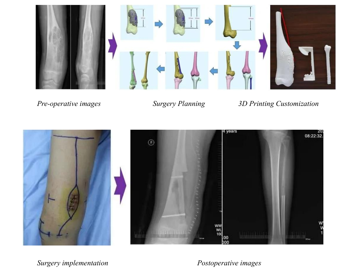
In this case, the patient is a 14-year-old adolescent. Taking into account the age and growth characteristics, combined with the location, size and shape of the tumor lesion, it was finally decided to use the proximal fibula graft with the donor site to preserve the epiphysis for autologous bone bio-reconstruction to repair the bone defect. However, this treatment protocol has no precedent in the world, so how to elaborately plan the surgical procedure from scratch, how to ensure the accurate implementation of the operation, how to guarantee the normal growth and development of the patient to the furthest extent, these were the challenges that the team had to solve.

Professor Liu Bin from DUT and Professor Shang Guanning from LCHI have conducted many technical discussions on the surgical plans and the repair and reconstruction methods. Finally, the research team used artificial intelligence technology to identify and extract lesions, and applied 3D printing technology for tailored autonomy bone biological reconstruction anatomy, biomechanics simulation matching, and precise simulation of surgery plan. With the assistance of Shenyang East Asia Orthopaedic Research Institute, the 3D printing model and the precise osteotomy guide plate design were completed, a thorough surgical plan was developed, and the operation was successfully completed. Six weeks after the operation, the patient’s autologous bone graft healed well, the fibula at the donor site was regenerated accurately, and he could walk with weight.
It is reported that in September 2020, DUT and LCHI signed the “DUT-LCHI comprehensive Cooperation Agreement” to jointly build a “medical-engineering joint research center” with its focus on big data precision medicine, biomedicine engineering, high-end medical equipment and other medical-engineering cross-cutting frontiers. They use their respective advantages to carry out scientific research cooperation and have achieved many results. This surgery is a successful attempt to apply scientific research results to medical practice.
In the future, DUT will vigorously expand the field of medical-engineering integration and further strengthen medical-engineering cross-cooperation to provide effective scientific and technological guarantees for people’s lives and health.